
Squamous Cell Carcinoma
Specialist assessment, diagnosis and treatment for squamous cell carcinoma in Birmingham
At Midland Skin we offer squamous cell carcinoma consultations with a Consultant Dermatologist for patients in Birmingham, Solihull, Coventry, Wolverhampton, Worcester and the Midlands.


Quick Facts about squamous cell carcinoma

Best for
Suspected or confirmed SCC

Appointment
Initial dermatology assessment

Treatment
Excision or Mohs surgery

Symptoms
Growing, crusted or tender lump

Provider
Consultant dermatologist

Price
See below
What is squamous cell carcinoma?
Squamous cell carcinoma, usually shortened to SCC, is the second most common form of skin cancer after basal cell carcinoma.
SCC develops from the keratinocytes that make up most of the outer layer of the skin. It is strongly associated with cumulative ultraviolet exposure from sunlight or sunbeds and most often appears on the face, ears, lips, scalp, backs of the hands, forearms and lower legs.
SCC often grows more quickly than BCC, sometimes over a few weeks or months. Unlike BCC, a small proportion of SCCs can spread to the lymph nodes or beyond. Most SCCs are cured by treatment, but the risk of spread and recurrence rises with size, depth, site and how long the tumour has been present. Early assessment allows the SCC to be treated while it is smaller and lower risk.
SCC can develop from a pre-existing sun-damaged patch (actinic keratosis) or from Bowen’s disease, which is an SCC confined to the surface layer of the skin.
Who is more likely to develop SCC?
People are more likely to develop SCC if they:
- have fair skin that burns easily
- have had significant sun exposure through outdoor work, outdoor activities, holidays or sunbeds
- have a weakened immune system, for example after an organ transplant, with certain blood conditions or on immunosuppressive medication
- have previously had an SCC, BCC, actinic keratoses or Bowen’s disease
- are older, although SCC can occur in younger adults
- have a long-standing scar, ulcer, burn or area of chronic inflammation
- have previously received radiotherapy or medical ultraviolet treatment
- have one of the rare inherited conditions associated with skin cancer
People of every skin colour can develop SCC. In darker skin, SCC more often arises on the lower legs, in scars or on skin that is not sun-exposed, and a lesion should not be dismissed because someone does not have the usual risk factors.
What does squamous cell carcinoma look like?
SCC does not have one single appearance. Common features include:
- a firm, scaly or crusted lump that keeps growing
- a dome-shaped lump that appears rapidly, sometimes with a central plug or crater
- a thickened, rough or wart-like growth
- a sore, ulcer or scab that bleeds and does not heal
- a persistent scaly patch that has become thicker, raised or tender
- on the lip, a persistent scaly, crusted or ulcerated area
SCC may be tender or painful, which is less usual for BCC. Some SCCs look very similar to a wart, an inflamed actinic keratosis, a keratoacanthoma or a BCC, and a persistent scaly patch may be mistaken for eczema or psoriasis. A face-to-face assessment is needed to establish the most likely diagnosis, and a biopsy is often required to confirm it.
When should I see a dermatologist?
Arrange an assessment if:
- you have a lump, scab or sore that has been present for more than a few weeks and is not healing
- a lesion is growing quickly, becoming thicker or crusting repeatedly
- a scaly patch has become raised, tender or has started bleeding
- a wart-like growth has appeared on sun-damaged skin
- a previously treated actinic keratosis or Bowen’s disease has changed or come back
- you have a weakened immune system or a previous skin cancer and notice any new or changing lesion
- your GP or another clinician suspects an SCC
- a biopsy has already confirmed SCC and you require treatment
- a previous excision has produced a close or involved margin
- you would like a second opinion about standard excision or Mohs surgery
Because SCC can grow and change over weeks, it is better to be assessed early than to wait and watch.

matthew stanley

How is squamous cell carcinoma diagnosed?
Your Consultant Dermatologist will examine the lesion and the surrounding skin, usually with a dermatoscope, and will check the nearby lymph nodes. Clinical photographs are taken as part of the assessment and treatment-planning record.
The dermatologist will consider:
- how long the lesion has been present and how quickly it has changed
- its size, thickness, position and how clearly its edges can be seen
- whether it is tender, ulcerated or fixed to deeper tissue
- previous skin cancers, sun damage or treatment in the same area
- your medical history, medication and immune status
In many cases a biopsy is recommended to confirm the diagnosis before definitive treatment is planned. For a small lesion, the biopsy may remove the whole lesion, but this cannot be assumed until the histology report is available. The histology report describes how deep the SCC extends and how well differentiated it is, which directly influences the treatment recommended.
Scans are not needed for most SCCs. Imaging may be recommended if the SCC has high-risk features or if there is any concern about the lymph nodes.
If your SCC has already been diagnosed
You can arrange an assessment at Midland Skin if your SCC was diagnosed by another private clinic or through the NHS.
Please provide your histology report, relevant clinic letters and any available photographs before the appointment. Your Consultant may also need to obtain or review additional pathology information before treatment is confirmed.
The original biopsy site can become difficult to identify once it has healed. Clear information about the precise location is therefore important.
What determines which SCC treatment is recommended?
SCCs are grouped into lower-risk and higher-risk tumours. This grading is the main factor in deciding the treatment margin, the surgical technique, whether specialist multidisciplinary discussion is needed and how closely you are followed up afterwards.
Features that place an SCC in a higher-risk group include:
- a diameter of more than 2 cm
- a thickness of more than 4 mm or invasion into fat, muscle or bone
- a poorly differentiated or aggressive growth pattern on histology
- involvement of nerves or blood vessels on histology
- location on the ear, lip, temple, scalp or other high-risk site
- a tumour that has come back after previous treatment
- an SCC arising in a scar, ulcer, burn or previously irradiated skin
- a weakened immune system
- poorly defined edges
A small, well-defined, well-differentiated SCC on the body is usually treated with standard surgical excision. A larger SCC, one in a high-risk site, one with poorly defined edges or one that has returned may be better treated with Mohs surgery or a wider excision, and may need multidisciplinary review. Your general health, preferences and ability to manage recovery are also taken into account.
“Higher risk” refers mainly to the likelihood of incomplete removal, recurrence or spread. It does not mean the SCC cannot be cured. Most higher-risk SCCs are still cured with appropriate surgery.
Your SCC treatment pathway
Initial dermatology assessment
You meet with a Consultant Dermatologist or Consultant Surgeon who examines the lesion and lymph nodes, reviews any previous biopsy or treatment and assesses the factors that influence diagnosis, tumour risk, treatment and reconstruction.
Biopsy or diagnostic review
A biopsy is recommended if the diagnosis or risk grade needs confirmation. If the SCC has already been diagnosed elsewhere, your histology report and other relevant records are reviewed.
Treatment plan
Your Consultant explains the recommended treatment, available alternatives, expected scar, recovery, risks and likelihood of clearance. Higher-risk SCCs may be discussed with specialist colleagues before the plan is finalised.
Treatment
Treatment usually involves standard surgical excision or Mohs surgery. Radiotherapy or other specialist treatment is arranged by referral where appropriate.
Results and follow-up
The histology result is reviewed with you. Follow-up is arranged according to the risk grade of the SCC, the treatment margins and your individual risk factors.
How is squamous cell carcinoma treated at Midland Skin?
The aim of treatment is to remove the SCC completely with a clear margin, confirm this on histology and repair the wound appropriately.
Standard surgical excision
Standard surgical excision is the main treatment for most SCCs.
The area is numbed using a local anaesthetic. The SCC is removed together with a planned safety margin of surrounding skin. Because SCC can extend beyond its visible edge, the margin is usually wider than for a BCC and is wider again for higher-risk tumours. The specimen is sent to a pathology laboratory so that the diagnosis, depth, differentiation and excision margins can be examined under a microscope.
Depending on the size and position of the wound, it may be closed directly with stitches, repaired with a skin flap or skin graft, or allowed to heal naturally.
If the histology shows SCC at or very close to an edge, further treatment is usually required.
Mohs micrographic surgery
Mohs micrographic surgery may be recommended for selectedSCCs, particularly where the edges are poorly defined, the tumour is on the face, ear or another site where preserving healthy tissue matters, or the SCC has returned after previous treatment.
During Mohs surgery the SCC is removed in stages. Each stage is mapped, processed in Midland Skin’s on-site frozen-section laboratory and examined under a microscope while you wait. If tumour remains at an edge, a further layer is removed only from that area until the margins are clear. This allows clearance to be confirmed before the wound is repaired.
Radiotherapy
Radiotherapy may be considered for selected SCCs where surgery is not appropriate or is declined, and is sometimes recommended after surgery for high-risk tumours. It is delivered by a specialist oncology team. Midland Skin can assess whether radiotherapy should be considered and arrange an appropriate referral.
Treatments that are not suitable for invasive SCC
Prescription creams such as imiquimod or 5-fluorouracil, cryotherapy and photodynamic therapy are used for actinic keratoses and Bowen’s disease. They are not appropriate treatments for an invasive SCC, because they do not remove the tumour reliably and do not allow the margins to be checked. If a lesion previously treated with a cream or freezing has persisted or returned, it should be reassessed.
Advanced or spreading SCC
A small number of SCCs spread to the lymph nodes or beyond. These are managed by a specialist skin cancer multidisciplinary team and may involve lymph node surgery, radiotherapy or immunotherapy. If your Consultant has any concern about spread, this is discussed with you and the appropriate referral is arranged.
Important limitations and risks
Every form of SCC surgery leaves a scar. The wound and scar are usually larger than the visible lesion because a treatment margin is required and the SCC may extend further beneath the skin than it appears on the surface.
Surgical risks include discomfort, bleeding, infection, wound-healing problems, altered sensation, an unsatisfactory scar and recurrence. A flap, graft or staged reconstruction may be required.
No treatment can guarantee that an SCC will never return or spread. Histology can occasionally show that the diagnosis, depth or risk grade differs from what was expected, which may change the treatment plan, require further surgery or lead to a recommendation for specialist multidisciplinary review.
Why choose Midland Skin for SCC treatment?
Midland Skin provides a consultant-led pathway from assessment and diagnosis through to treatment and follow-up.
- Suspicious lesions and confirmed SCCs are assessed by Consultant Dermatologists with skin cancer surgery experience.
- Standard surgical excision and Mohs surgery are both available, so the technique is chosen according to the risk grade of the SCC.
- Mohs surgery is supported by an on-site frozen-section laboratory.
- You meet the clinician accepting responsibility for your procedure before treatment.
- Wounds can be repaired using direct closure, skin flaps, skin grafts or natural healing, depending on the defect.
- Specialist reconstructive and oncology colleagues can be involved, and higher-risk SCCs can be referred for multidisciplinary discussion where appropriate.
- Care is provided within a CQC-registered Birmingham clinic.
Can squamous cell carcinoma be cured and can it return?
Most SCCs are cured by complete surgical removal. The outlook is best when the SCC is treated while it is small and thin.
An SCC can occasionally return in the same place after treatment, and a small proportion spread to the lymph nodes. The great majority of recurrences and spread occur in higher-risk tumours and within the first two to three years after treatment, which is why follow-up is structured according to risk.
- A completely excised lower-risk SCC may need little or no routine clinical follow-up beyond a wound review and advice on self-checking.
- A higher-risk SCC is usually followed up at regular intervals for several years, including examination of the treated area and lymph nodes. Follow-up may be arranged at Midland Skin or, where appropriate, through an NHS skin cancer service.
Recurrence at the treatment site is different from developing a completely new skin cancer elsewhere. Once you have had one SCC, your risk of further SCCs, BCCs and actinic keratoses is increased, particularly if your immune system is suppressed.
You should check your skin approximately once a month, including the treated area and any nearby lymph nodes, and arrange an assessment if you notice a new or changing lump, scab, patch or non-healing area.
Protecting the skin from ultraviolet exposure helps reduce further damage. This includes avoiding sunbeds, avoiding sunburn, using protective clothing and applying a broad-spectrum sunscreen to exposed skin. Treating actinic keratoses and Bowen’s disease early reduces the chance of a further SCC developing.
John Osborn
SCC assessment and treatment: FAQs
I already have a confirmed SCC. Can Midland Skin treat it?
Yes. We assess patients whose SCC has been diagnosed through the NHS or by another private provider. Please send us your biopsy report, relevant clinic letters and any available photographs. Your Consultant will review the diagnosis, risk grade, treatment options and likely reconstruction before agreeing the procedure.
Why do I need an assessment when I already know I have an SCC?
The clinician accepting responsibility for your procedure must assess and plan it properly. This includes confirming the diagnosis and treatment site, reviewing the histology and risk grade, examining the lymph nodes, assessing the visible and possible microscopic extent of the SCC, deciding on the appropriate margin or surgical technique and planning how the wound will be repaired.
Your medical history, medication, bleeding risk, healing risk and treatment preferences must also be considered. This assessment is a separate professional service from the procedure and its fee is not deducted from the treatment fee. There is no obligation to proceed with treatment at Midland Skin.
Can the SCC be removed at my first appointment?
Assessment and SCC treatment are normally arranged as separate appointments. You should not assume that a biopsy or definitive removal will be carried out during the initial assessment. This allows the diagnosis, surgical margin, histology requirements, procedure time and wound repair to be planned properly.
Will I need a biopsy?
Often, yes. Confirming the diagnosis and risk grade before definitive surgery allows the correct margin and technique to be chosen. A biopsy may not be needed if the appearance is characteristic and the lesion can be removed as a complete excision with an appropriate margin. Your Consultant will explain which approach is recommended.
Can an SCC be treated with a cream or by freezing instead of surgery?
No. Creams, cryotherapy and photodynamic therapy are used for actinic keratoses and Bowen’s disease, not for invasive SCC. If a lesion that was treated in this way has persisted or returned, it needs reassessment and may require a biopsy.
How soon can I be assessed and treated?
Initial consultations are usually available within two to three weeks. Once treatment has been agreed, surgery is generally arranged within the following weeks, depending on clinic availability, the urgency of the SCC and your preferences.
What happens if the excision margins are not clear or the SCC is high risk?
Further treatment is usually required when SCC is present at an excision margin. Options may include further excision, Mohs surgery or radiotherapy, depending on the tumour and its site. Higher-risk SCCs may be discussed at a specialist skin cancer multidisciplinary team meeting before the next step is agreed, and your Consultant will explain what this involves.
Will SCC surgery leave a scar?
Yes. Any form of skin cancer surgery will leave a scar. The scar is usually longer than the visible SCC because a treatment margin is required and the wound must be repaired safely. Its final appearance depends on the size, depth and position of the SCC, the repair required and how your skin heals.
Can several lesions or a full skin check be included in the same assessment?
The specialist skin surgery assessment is arranged for the lesion or lesions agreed when you book. If you have several lesions, extensive sun damage or would also like a wider skin examination, please tell the office before booking so that the correct appointment type and sufficient time can be arranged.
Book in with us
Ready to get started? Book a consultation with our team at Midland Skin. Friendly, expert advice tailored to you.
Fees for SCC assessment and treatment
SCC Show pricing
Book ConsultationThe consultation fee covers the consultant’s assessment, examination, clinical photographs, clinical judgement and surgical care plan. It remains payable whether surgery, another treatment or no treatment is recommended. It is separate from, and is not deducted from, the fee for any subsequent procedure. If surgery is recommended, the procedure fee will be confirmed once the required treatment has been established. Fees for surgery include any immediate aftercare that is required, such as wound checks, dressing changes and stitch removal.
Initial dermatology assessment
Up to 25 minutes
Biopsy of skin
Skin cancer excision (per lesion)
Skin cancer excision plus flap or skin graft
Mohs surgery for skin cancer
Includes first review consultation
References used for this article
- British Association of Dermatologists. Squamous cell carcinoma patient information leaflet.
- Keohane SG, et al. British Association of Dermatologists guidelines for the management of people with cutaneous squamous cell carcinoma 2020. Br J Dermatol. 2021;184(3):401–414. doi:10.1111/bjd.19621.
- Stratigos AJ, et al. European consensus-based interdisciplinary guideline for invasive cutaneous squamous cell carcinoma: part 2. Treatment – update 2023. Eur J Cancer. 2023;193:113252.
- Tomás-Velázquez A, et al. Risk factors and rate of recurrence after Mohs surgery in basal cell and squamous cell carcinomas: a nationwide prospective cohort. Acta Derm Venereol. 2021;101(11):adv00602. doi:10.2340/actadv.v101.544.
About the Author
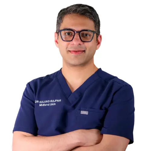
This page has been written and/or medically reviewed by Dr Sajjad Rajpar, Consultant Dermatologist and Medical Director of Midland Skin, Birmingham.
Dr Rajpar is on the GMC Specialist Register in Dermatology and has over 20 years’ experience in clinical dermatology. He provides clinical oversight to ensure the information on this page is accurate, balanced and consistent with current dermatology practice.
Dr Sajjad Rajpar
Date last updated: 26th Sep 2026
Book in with us
Expert, consultant led care in dermatology, skin surgery, eyelid surgery, lasers and aesthetics.
Disclaimer
The content on this website is provided for general information only. It is not, and must not be treated as, medical advice, a diagnosis, or a recommendation for any particular treatment. We make reasonable efforts to keep information accurate and up to date, but medicine and technology change, and content may not reflect the most current evidence, guidance, product information, or individual practice. You should not rely on this website to make decisions about your health or treatment. Always seek personalised advice from an appropriately qualified healthcare professional. Any examples, images, testimonials, and outcome descriptions are illustrative only. Results vary between individuals and no outcome can be guaranteed.


